5028 INTRALOBAR SEQUESTRATION
The patient was a male aged 11 years, and had had lobar pneumonia after whooping cough when 18 months old. It cleared without incident, but nine years later the pneumonia recurred and he was found to have a large cystic opacity at the left base. A lobectomy was performed and a large multicystic mass was found occupying most of the left lower lobe. A large aberrant artery was found running from the aorta through the lower border of the pulmonary ligament to the cystic mass.
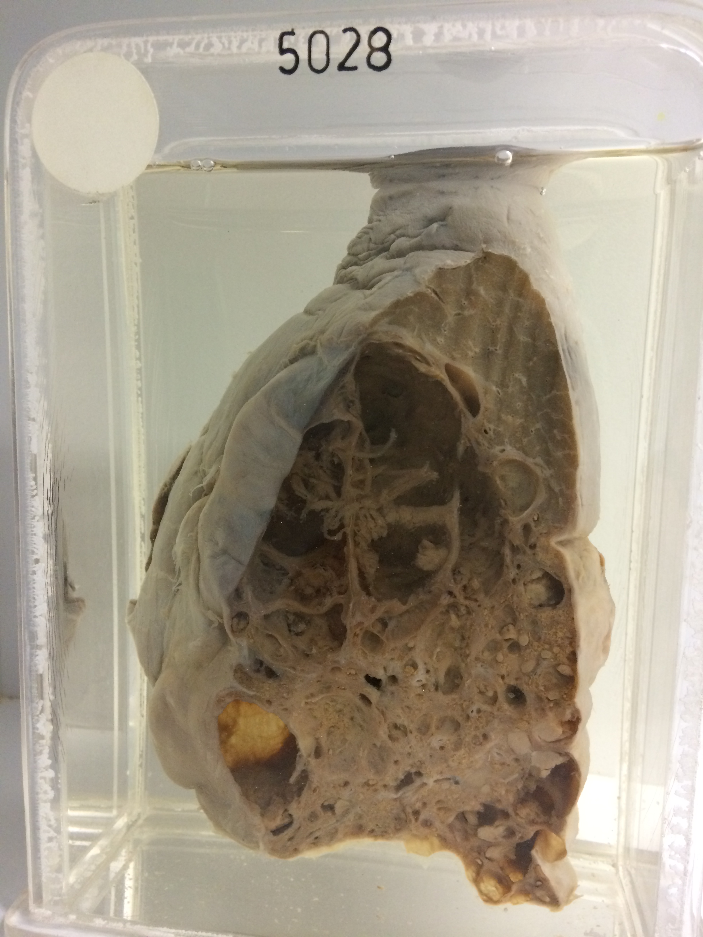
The specimen consists of the lower lobe of the lung sectioned to show a large mass of cystic cavities replacing the pulmonary tissue. Thick fibrous strands traverse the cavities and many of the smaller spaces are filled with purulent exudate. There are other small areas of lipid pneumonitis. The adjacent lung is displaced but appears essentially normal. A large abnormal artery arising from the aorta to supply this malformation is visible on the reverse of the specimen entering the posterior basal segment of the lower lobe. Histology shows cysts lined with cubical to columnar epithelium. There are many foamy macrophages in the areas of lipid pneumonitis and there is purulent exudate in the infected cysts.