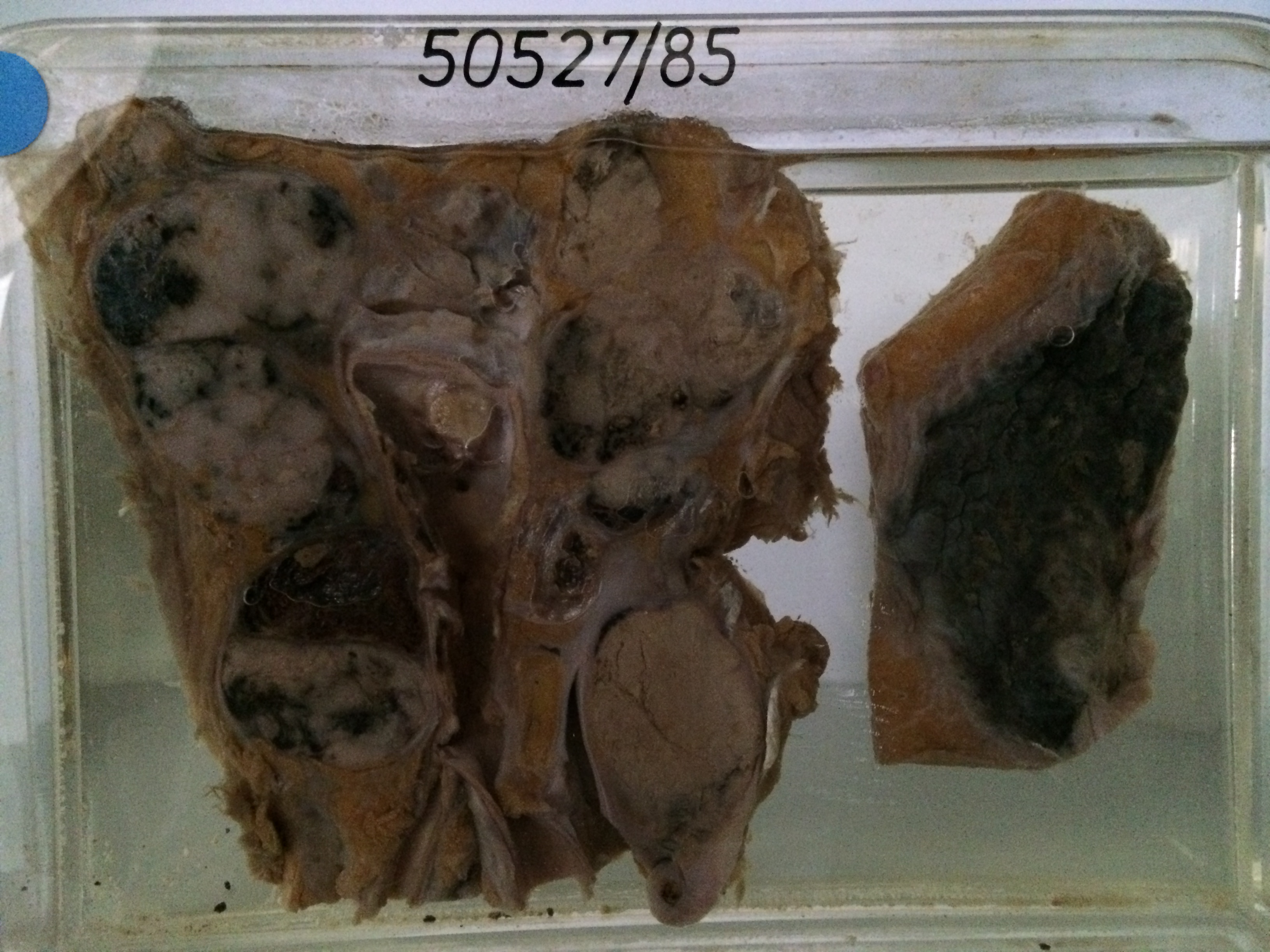
50527/85 ADENOCARCINOMA OF PROSTATE
The specimen on the right side includes a segment of bladder with prostate. When looked at from below it can be seen that white tumour tissue has destroyed the normal architecture of the prostate and extends out into the surrounding fat. One seminal vesicle is included in the slice. The bladder mucosa is oedematous, congested and in places haemorrhagic reflecting cystitis. The larger specimen is aorta, which is atheromatous, and the adjacent inferior vena cava. These vascular structures are surrounded by numerous lymph nodes which are enlarged and replaced by metastatic tumour which is grey but in places is showing necrosis (yellow) and haemorrhage (red or black).
Last modified: Monday, 31 July 2017, 11:46 AM