9510 INTERSTITIAL PULMONARY FIBROSIS
This patient was a woman aged 59 who had had chronic idiopathic pulmonary fibrosis for 6 years. She was admitted to hospital on many occasions with respiratory infections. She gradually became more breathless, with a productive cough and at her final admission there was tachycardia, the B.P. was 95/75 and the vital capacity was 500 mls. She died on the following day. At postmortem there was mild finger clubbing and some nephrosclerosis. The liver was reported as congested but the heart weight only 260 gms and the right ventricle was not reported to be enlarged.
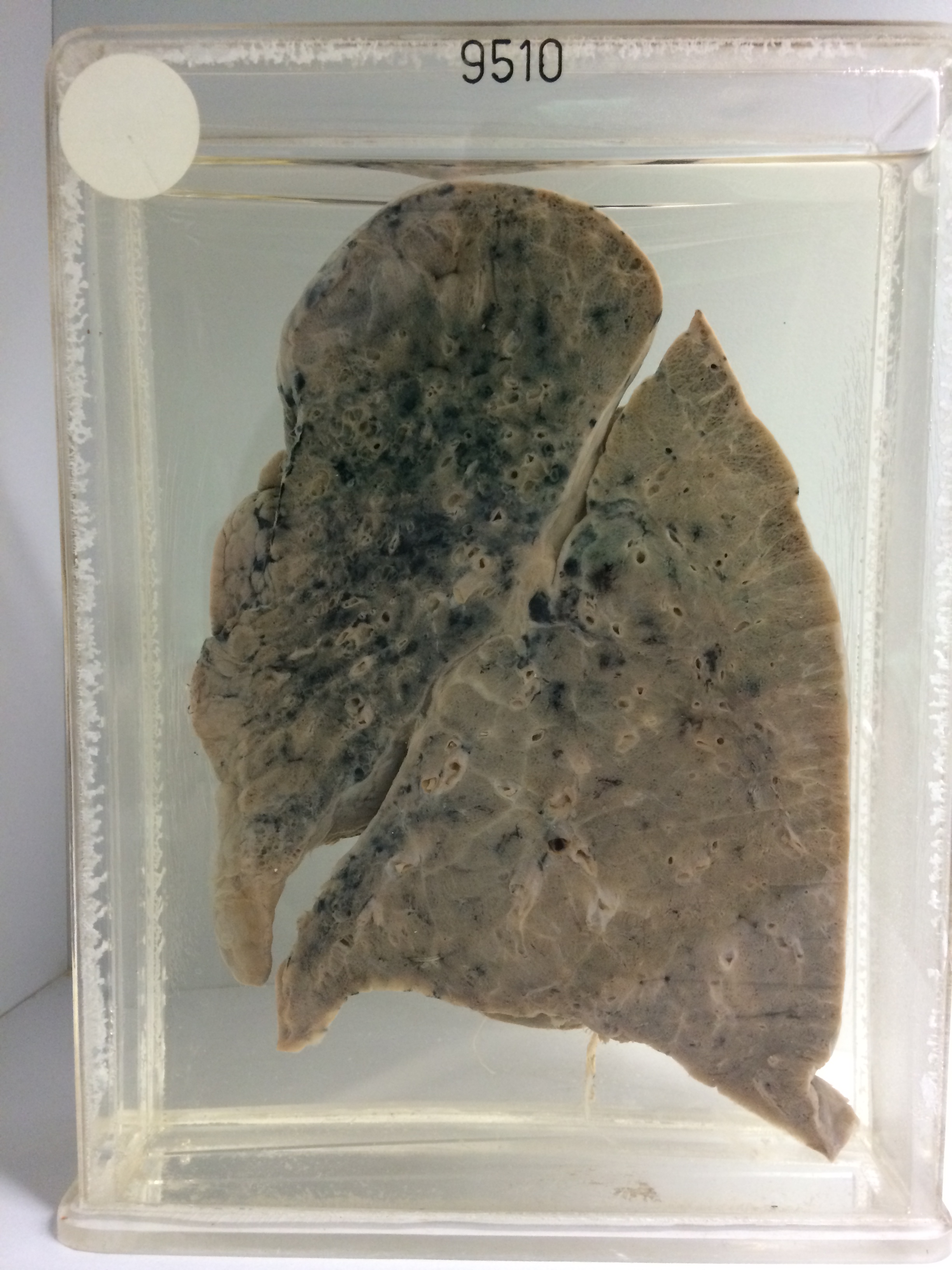
The specimen is the sectioned left lung which shows a diffuse interstitial fibrosis in the hilar aspect of the upper lobe extending in reticular fashion towards the surface. There are some thinwalled cystic spaces about 3 mms in diameter at the periphery of the fibrotic area. Antemortem thrombus is visible in a small artery in the lower lobe. Histology shows marked nodular and diffuse interstitial fibrosis. Chronic inflammatory cells are numerous. There is bronchialisation of epithelium in the cysts. Foreign body giant cells are quite numerous around cholesterol crystals in the stroma and around masses of what appears to be degenerating elastic tissue. Some foamy macrophages are present in some of the spaces.