20196 OLIGODENDROGLIOMA OF THE RIGHT FRONTAL LOBE
A woman aged 59 had been referred to a mental hospital with a diagnosis of presenile dementia. Apparently her mental faculties had been deteriorating for 5 years until she had become unable to feed herself. Examination showed chronic papilloedema, bilateral grasp reflexes, poor visual acuity but brisk light reflexes. Skull X-ray showed erosion of the dorsum sellae in keeping with raised intracranial pressure and there was an area of calcification in the right frontal lobe suggestive of tumour. She was accordingly transferred to the R.A.H. where a burr hole biopsy was performed which showed the frontal tumour to be a well differentiated oligodendroglioma. After the operation she developed a respiratory infection and died about 2 weeks later.
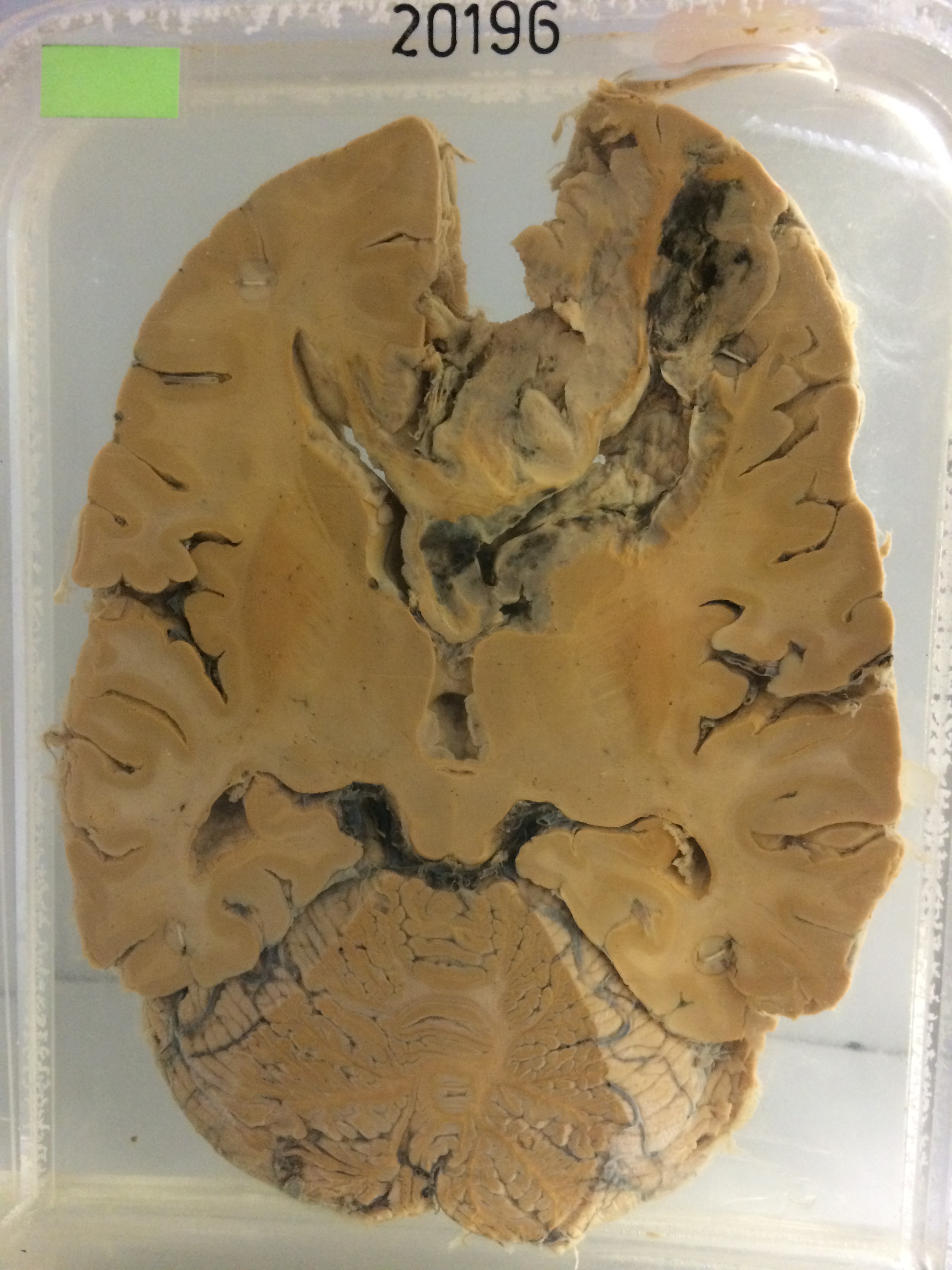
The specimen consists of a horizontal slice of the hemispheres together with the brain stem. There is a cellular tumour on the medial aspect of the right frontal lobe which extends across the genu of the corpus callosum for a little distance into the left frontal lobe. A large cavity lined with necrotic tissue and blood clot lies posterior to the tumour. There is some dilation of the 3rd ventricle and the ependymal lining of the anterior horn of the left lateral ventricle is very nodular. Histology shows an oligoglioma which is slightly more pleomorphic than usual.