12200 PAPILLARY CARCINOMA OF THE URETER
The patient was a woman aged 66 who had painless haematuria for 5 months. There was no dysuria or frequency. IVP showed a nonfunctioning right kidney. A block was demonstrated in the right ureter by contrast radiography. The kidney and ureter were removed surgically.
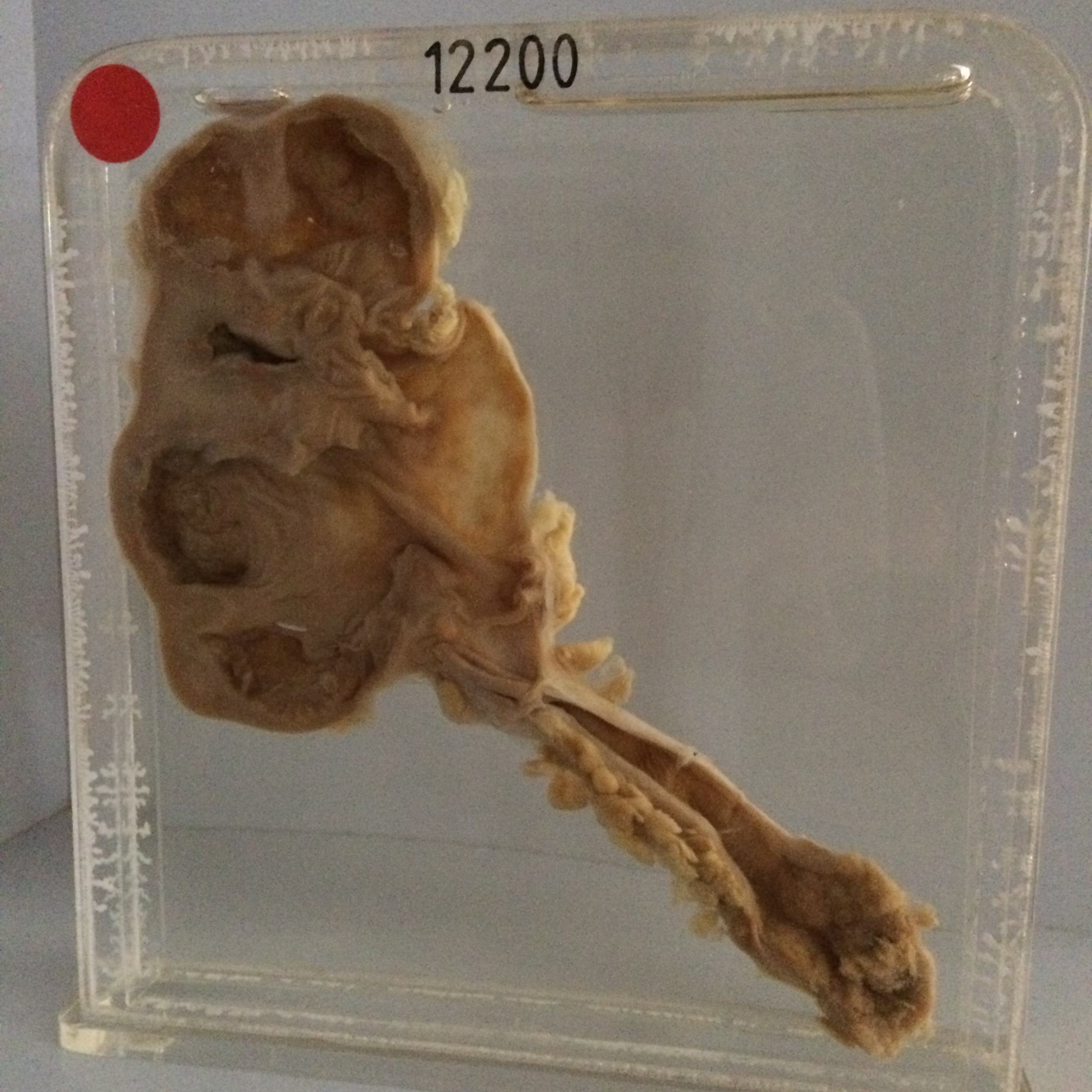
The specimen consists of a coronal slice of the kidney which measures 10 cms in length together with the upper 9 cms of the ureter. A fungating papillary carcinoma 2.5 cms in length blocks the ureter at a point 7 cms below its origin. Above the tumour the ureteric muscle is obviously hypertrophied but the lumen is only moderately dilated. The kidney shows gross intrarenal and moderate extrarenal hydronephrosis with a relatively narrow rim of renal tissue remaining. There is considerable shaggy material on the inner surfaces of the dilated calyces. Histology shows the ureteric tumour to consist of large irregular carcinomatous cells with much variation in size, shape and staining. There is some squamous metaplasia. Mitotic figures are numerous. The kidney is densely fibrous with many hyaline glomeruli. The 'exudate' on the inner surfaces of the dilated renal calyces consists of a diffuse layer of papillary carcinoma. This specimen therefore shows multiple foci of tumour.