12849 OSSIFIED SUBDURAL HAEMATOMA
The patient was a jockey aged 28. Seven months before his death he fell from his horse and sustained severe head injuries. Various neurosurgical procedures were performed without much success and the patient was left a mental defective. He was transferred to a mental hospital where he died 3 months later. At postmortem 4 burr holes and an osteoplastic flap were present in the skull, with a residual bony defect measuring 9 x 9 cms. The left cerebral hemisphere was extensively atrophied particularly in the frontal and parietal lobes. The right cerebral hemisphere was less affected but the arachnoid was stained with yellow pigment. Overlying the right posterior parietal region was an extensive thickening of the skull.
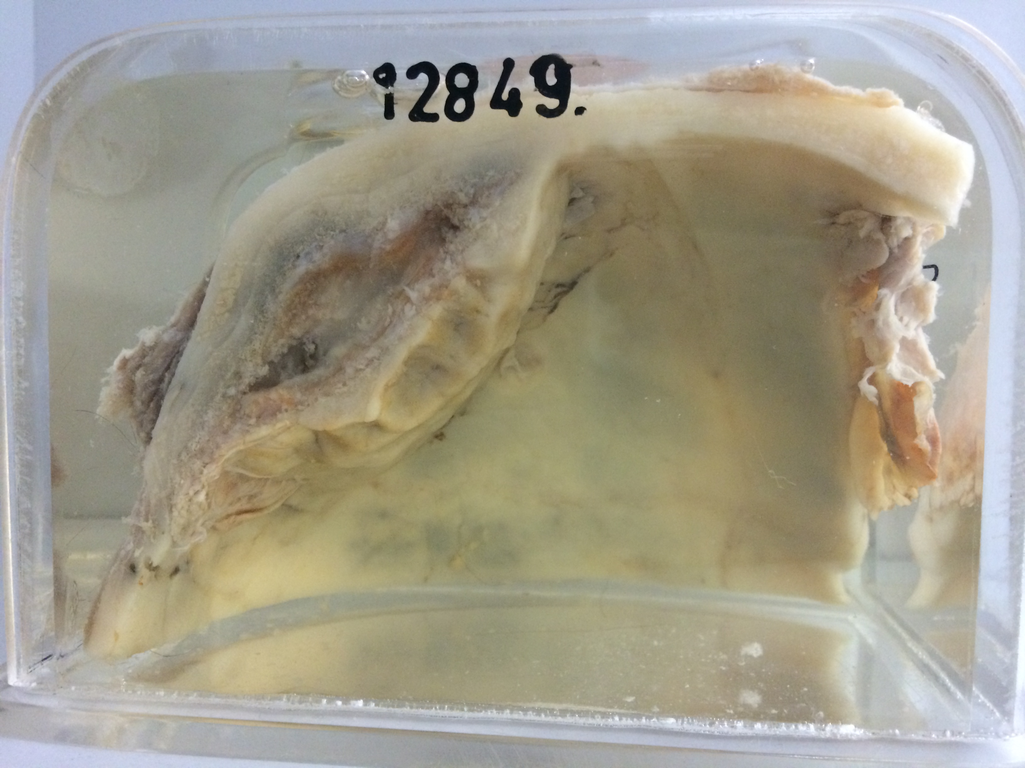
The specimen is of this portion of the skull divided in the coronal plane to reveal a large ossified subdural haematoma measuring 6 cms in diameter and 3 cms in thickness. The inner wall is of dense nodular bone and the cavity contains brown-stained fibrous tissue. Histology shows the centre to consist of loose oedematous fibrous tissue with scattered macrophages containing old blood pigment. The inner aspect is a layer of dense fibrous tissue which is also pigmented and within this is a thick layer of well differentiated cancellous bone.