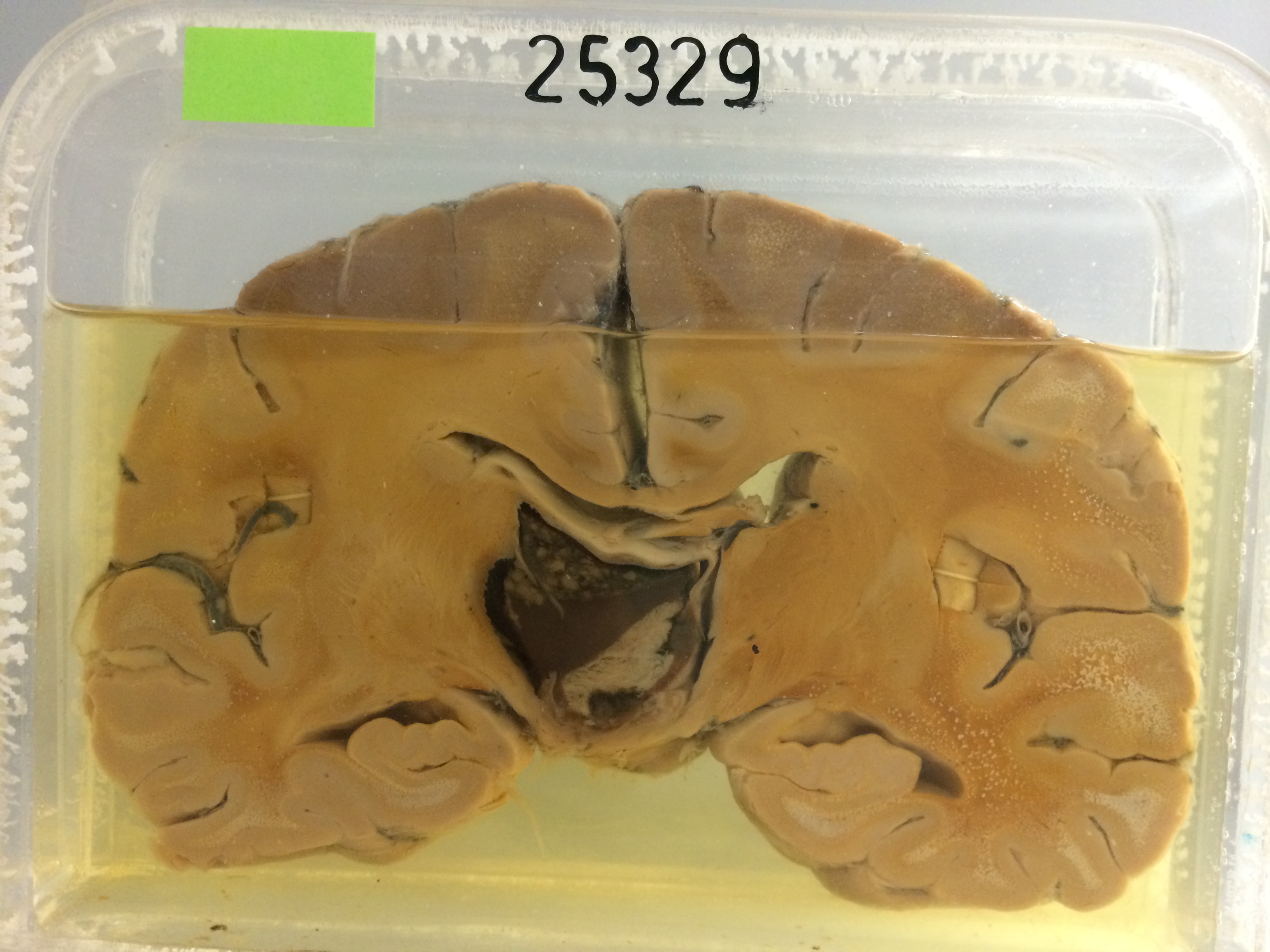
25329 COLLOID CYST OF THIRD VENTRICLE
The patient was a diabetic hypertensive woman aged 52. She was first admitted 5 months previously with sensory disturbance and right hemiparesis, dysphasia, and right homonymous hemianopia. Lumbar puncture showed xanthochromia and carotid angiography a shift of the pericallosal arteries to the right of the midline. Ventriculogram showed complete obstruction of both foramina of Munro. A left posterior frontal burr hole was made and altered blood was aspirated from the lesion. Thereafter her condition fluctuated, but she finally became comatose with fixed dilated pupils and died 2 days later.
The specimen consists of a slice through the hemispheres to disclose a unilocular cyst 2½ cms in diameter distending the septum pellucidum and attached to the left thalamus. The cyst contains clear gelatinous material with some more solid inclusions which appear cellular. Inferiorly there is a solid strip of grey tissue extending obliquely upwards and to the right, and the bottom of the specimen shows a very definite nodular bulging of the hypothalamus. Histology shows that the cyst contains strongly PAS-positive material. It is lined by an epithelium which is generally several layers thick, the cells varying from elongated forms through polygonal shapes to rounded cells. Many of the superficial cells are mucus-secreting. The more solid area inferiorly is a granulomatous mass containing many cholesterol clefts, between which is some hyaline fibrous tissue containing many red cells and macrophages, some filled with PAS-positive material, others with haemosiderin granules. It probably results from the settlement of haemorrhage, clot and debris to the bottom of the cyst.