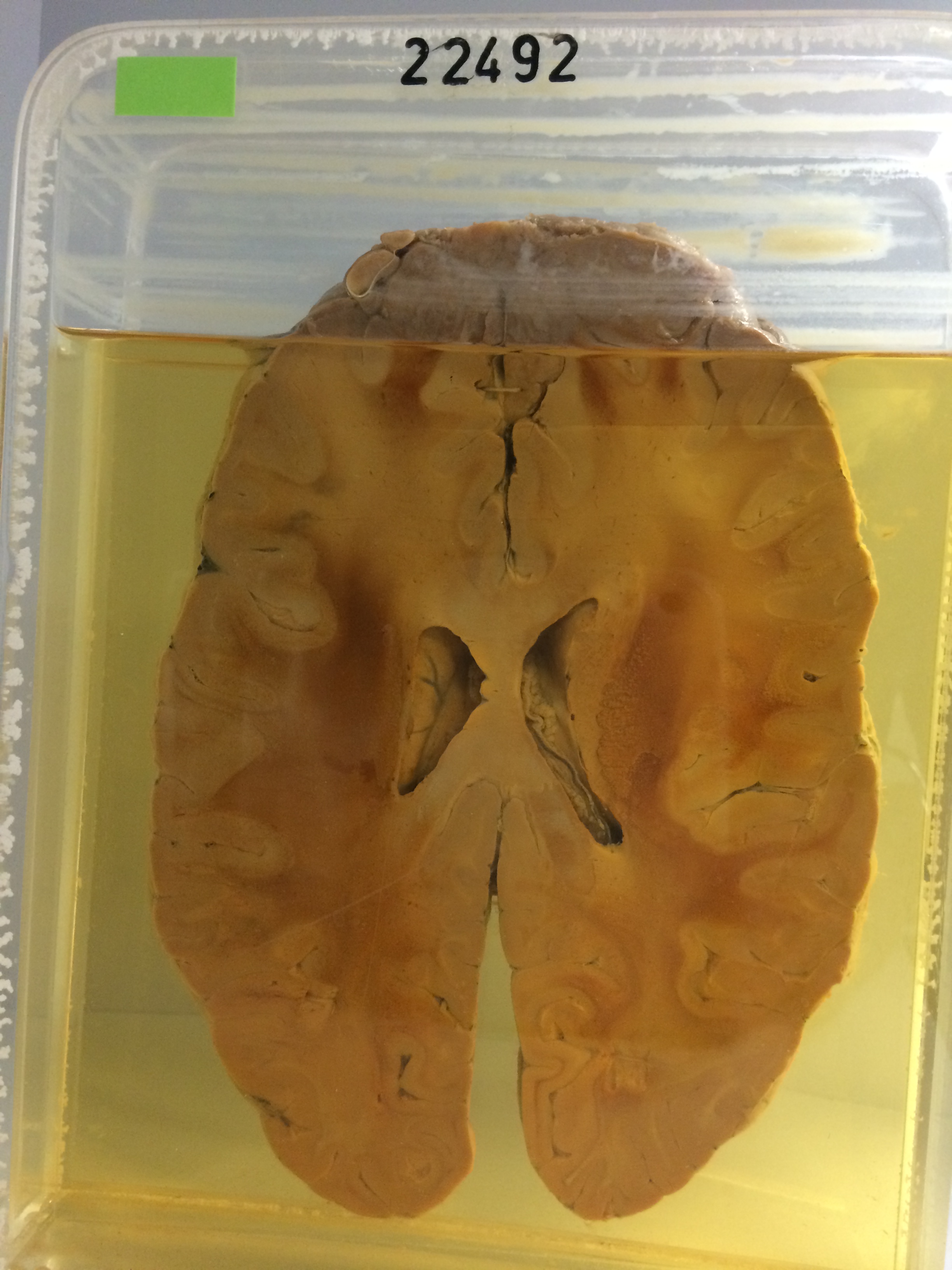
22492 HODGKIN’S LYMPHOMA
A man aged 20 had been treated for Hodgkin’s disease for 4 years, mostly by radiotherapy. At postmortem massive deposits of Hodgkin’s tissue were present in many regions, including the meninges.
The specimen is a horizontal slice through the cerebral hemispheres to disclose a large mass of pale Hodgkin’s tissue arising from the falx in the midline at the frontal poles. Posterior to this lymphomatous mass there is marked yellow oedema of the white matter of the frontal lobes. Both hemispheres are generally swollen and the cavities of the lateral ventricles are somewhat compressed. Histology shows anaplastic pleomorphic Hodgkin’s sarcoma with many Reed-Sternberg giant cell.
Last modified: Monday, 31 July 2017, 1:21 PM