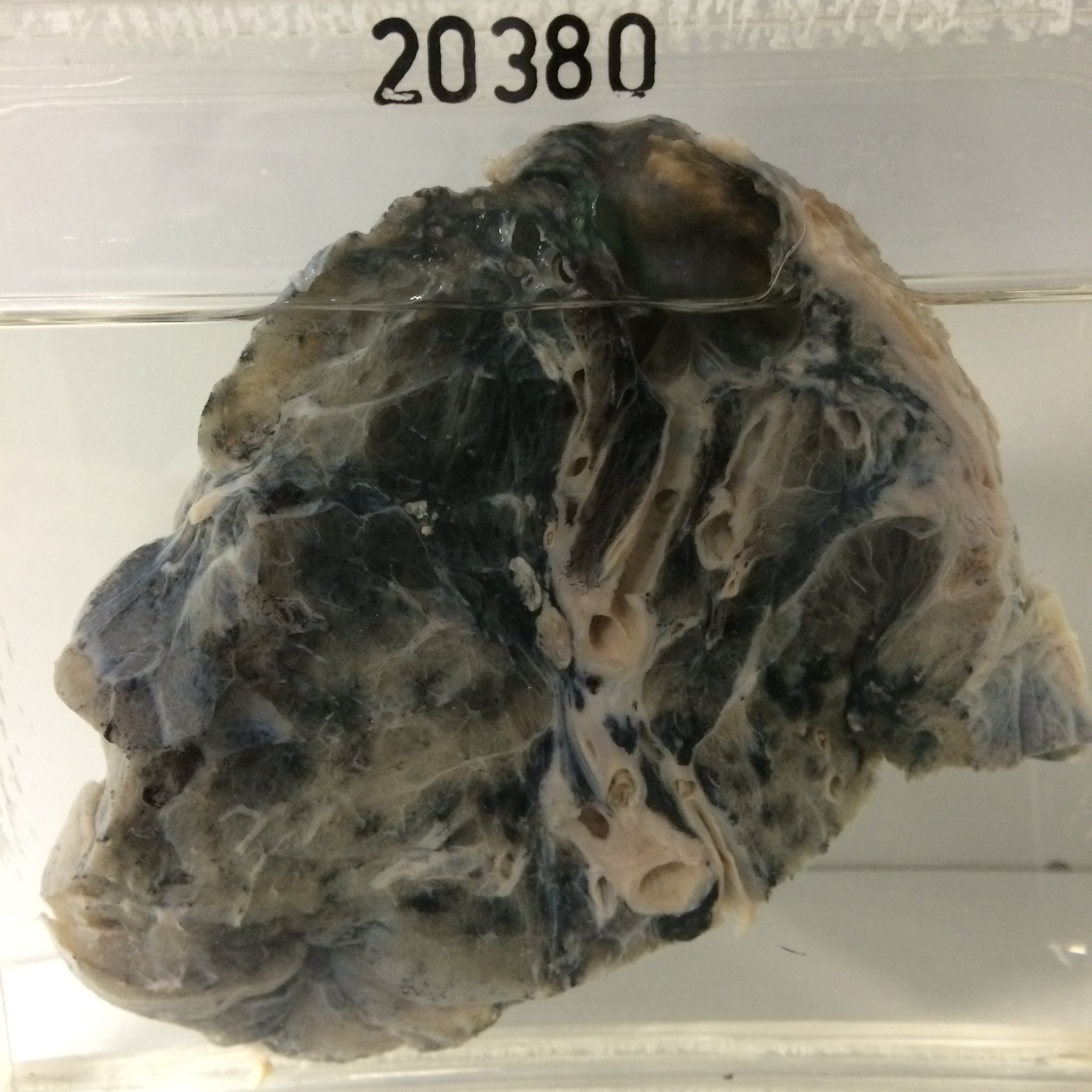
20380 HEALED TUBERCULOUS CAVITY WITH BRONCHIAL COMMUNICATION
This patient was a man aged 52. Eighteen months before his death he was found to have pulmonary tuberculosis with positive sputum and cultures. This was treated with streptomycin, PAS and INH and he slowly improved. Nine months later an increasing opacity was noticed in X-ray in the left mid-zone, consistent with a carcinoma. Malignant cells were not found in the sputum. Surgery was considered too great a risk because of his poor respiratory function. Six months later the mass in the lung had increased greatly in size and he died after a short time in hospital. At postmortem a massive carcinoma of the left lower lobe was found with metastases in the hilar nodes and in both adrenals. There was a cavity at the apex of the right lung.
The specimen is of the upper lobe of the right lung sectioned to show the thinwalled apical cavity 3 cms in diameter lying immediately beneath the pleura and surrounded by fibrotic and pigmented lung. A free communication with a bronchus is evident. There is marked destructive panlobular emphysema with anthracosis in the subapical pulmonary tissue and there is antemortem thrombus in medium-sized vessels. There is no evidence either grossly or histologically of residual active tuberculosis. The tumour in the other lung was a poorly differentiated adenocarcinoma.