25493 PEPTIC OESOPHAGEAL ULCER ATTACHED TO LEFT ATRIUM POST MORTEM ULCERATION
The patient was an old woman aged 86 who was admitted after a sudden very severe dull deep chest pain radiating into the neck. Seven years previously a hiatus hernia had been repaired and a year later there was an episode of melaena. Four years before her final admission she had a gastric ulcer and later pneumonia developed and a pericardial friction rub was heard. A diagnosis of mediastinitis due to oesophageal ulceration was accordingly made. On the 15th day, she died from a brain stem infarct. At post-mortem, the body was extensively decomposed, the liver was autolysed and there was bright red staining of the aorta. There was extensive surgical emphysema of the mediastinum, the retroperitoneal mesentery and the anterior chest wall and neck.
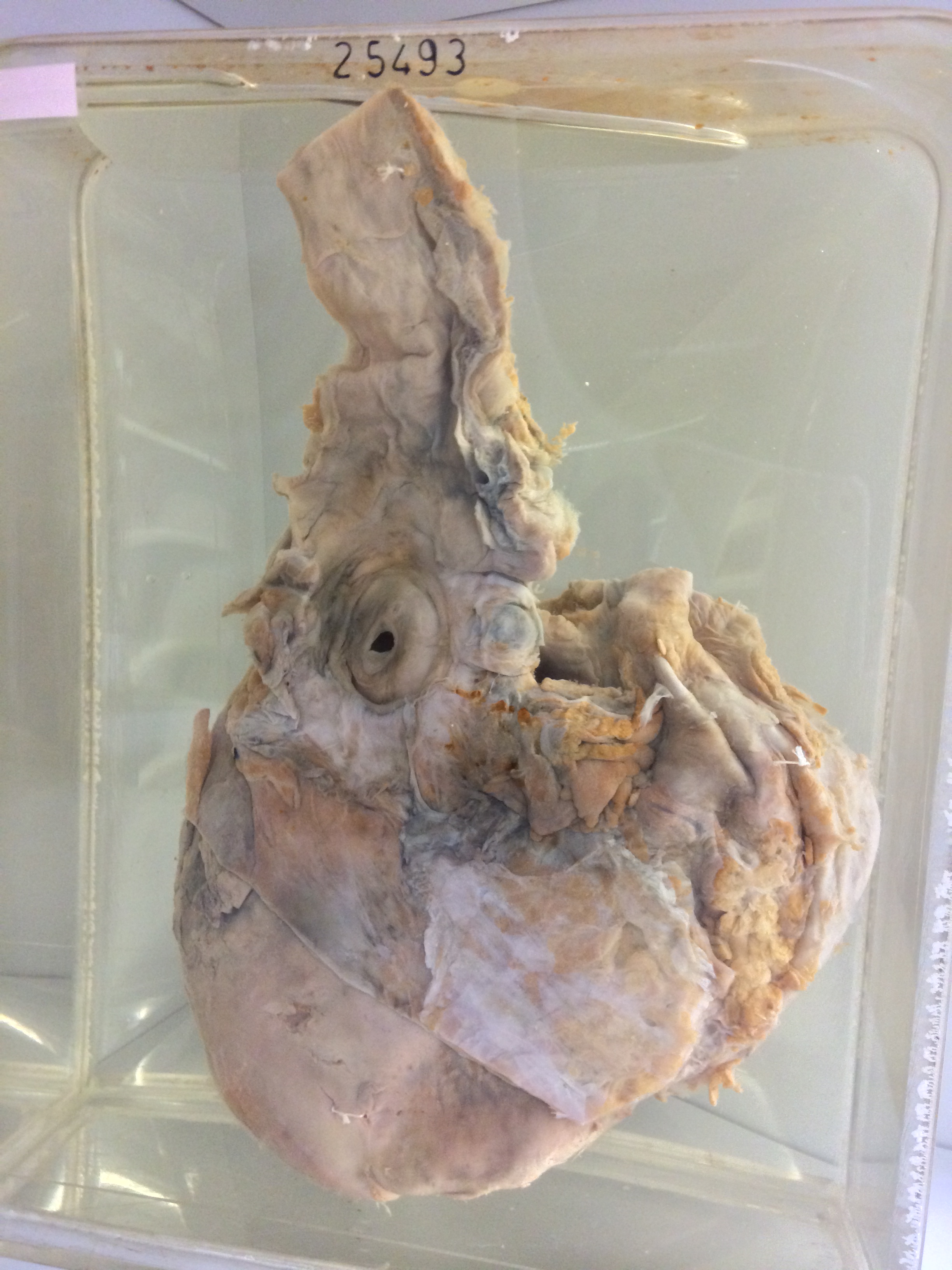
The specimen consists of the lower oesophagus together with the heart. A large superficial ulcer 2.5cm in diameter is present at the lower end of the oesophagus. It has regular margins and a smooth floor and there is a central perforation measuring 5mm x 3mm which leads into the cavity of the left atrium. The left and right ventricles are both dilated and hypertrophied. There is no record in the history likely that the perforation is artefactual due to post-mortem digestion. It seems likely that the ulcer became adherent to the wall of the atrium and it is possible that during life, there was some leakage from it into the mediastinum but not into the atrium.