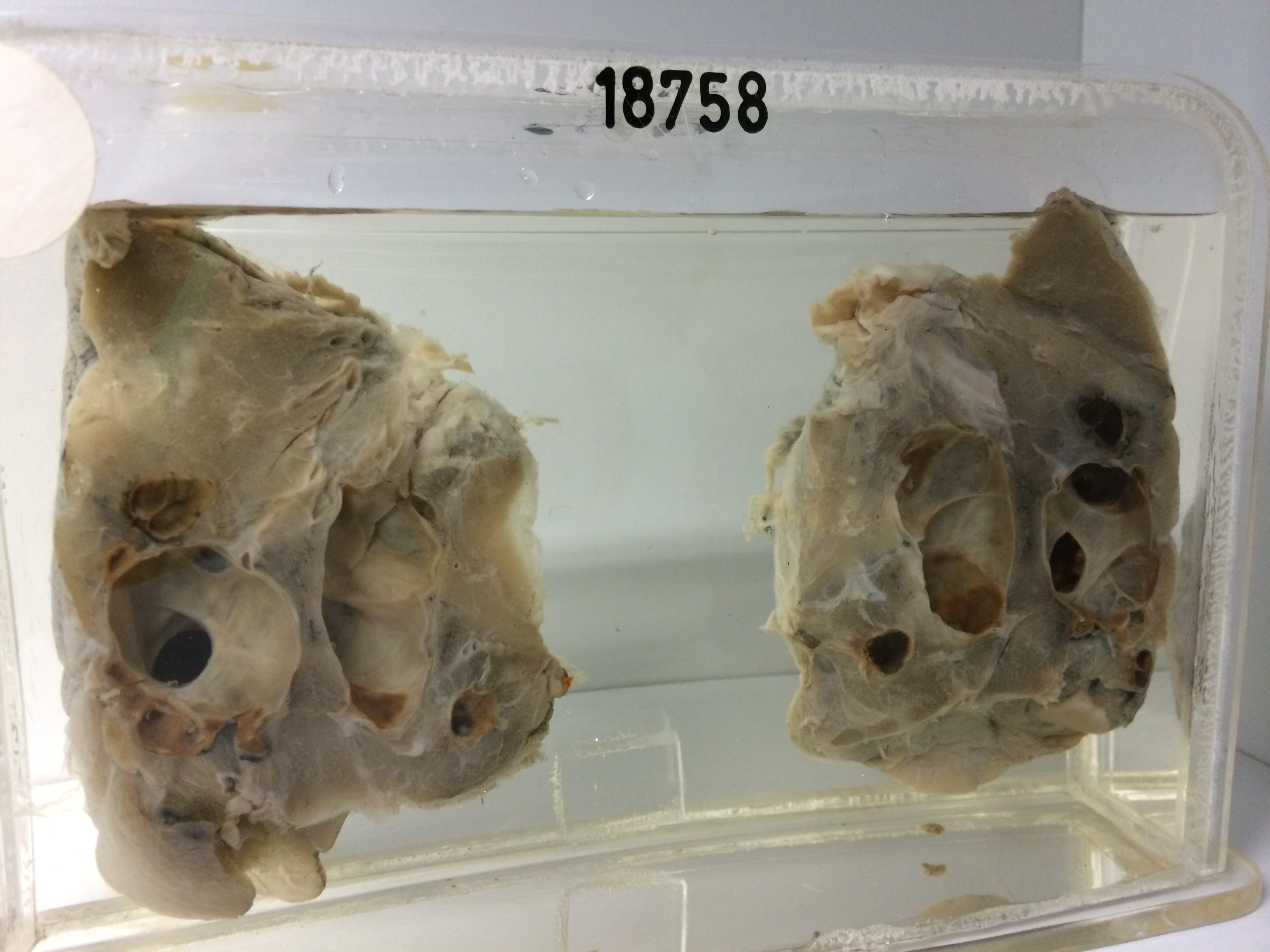
18758 CONGENITAL CYSTS OF THE LUNG
The patient was a woman aged 29 who had no symptoms. A lesion was discovered in the left lower lobe on routine chest X-ray. Review of earlier chest films then showed that the lesion had been present 4 years previously. The left lower lobe was resected and the specimen consists of this lobe bisected to show a collection of thin walled intercommunicating cysts with large openings between the cysts. The cyst lining is smooth. The intervening lung is a little fibrous but not obviously infected. There are some local areas of dense white fibrosis in the overlying pleura but no aberrant vessels supply this abnormality and no bronchial communication can be found by probing. Histology shows cysts lined by tall columnar ciliated epithelium which is missing in many places and is replaced by macrophages containing iron pigment. The intervening lung is fibrotic and partly collapsed.