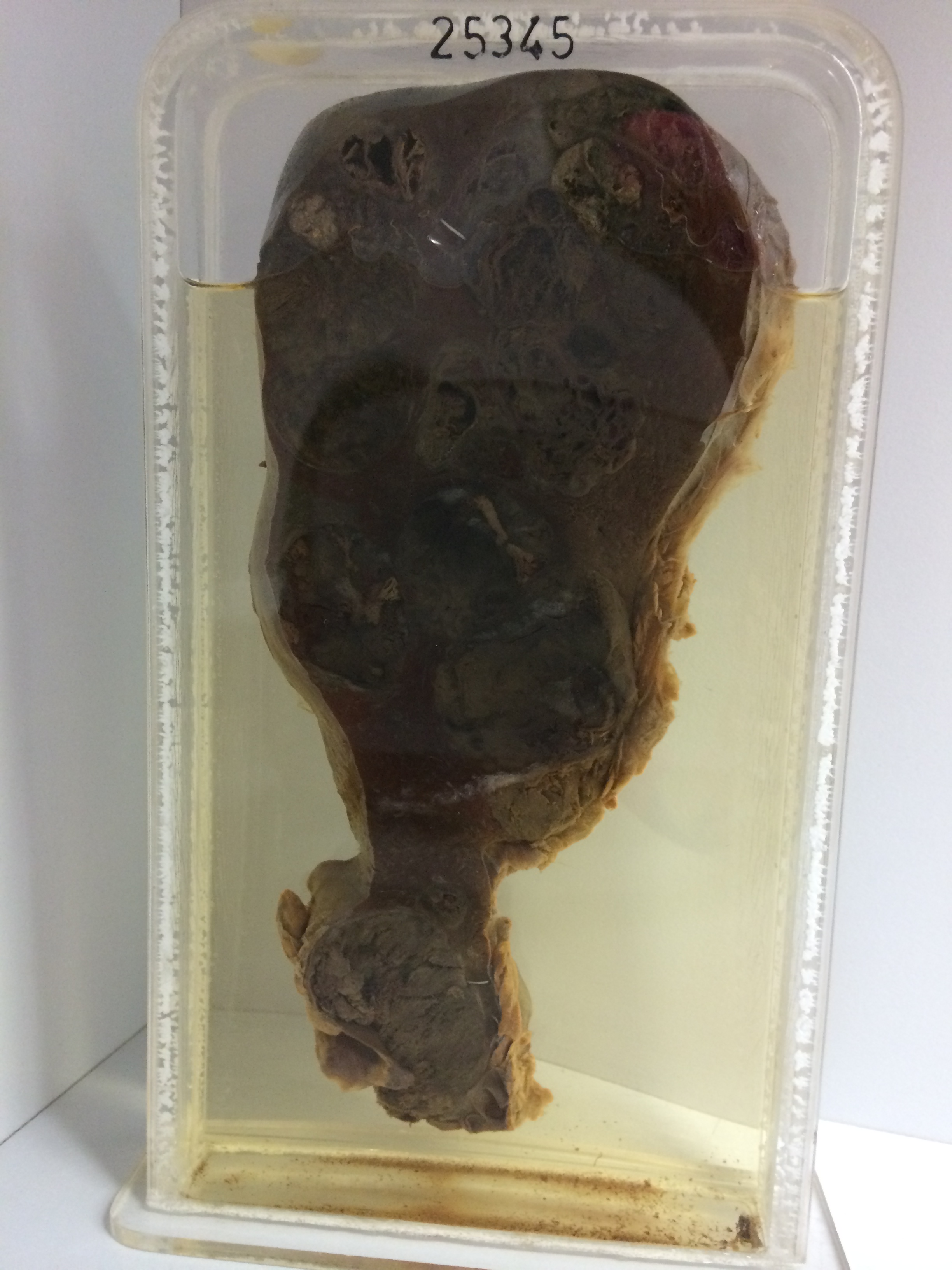
25345 SECONDARY MALIGNANT MELANOMA
The patient was a woman aged 54. A malignant melanoma had been excised from the left popliteal region a year previously. Four months later block dissection of the left inguinal nodes was performed, and after a further 4 months radiotherapy was given to the pelvic region. Two months later cytotoxic drugs were given. At this time chest X-rays showed metastases in both lungs. Cytotoxic drugs and steroids were continued, but she went steadily downhill and died. At postmortem widespread metastatic deposits were present in both lungs, the myocardium of the left ventricle, the kidneys, liver and spleen.
The specimen consists of a longitudinal slice through the enlarged liver. It measures 22 x 8 cms. There are numerous metastatic deposits throughout the liver substance. Some portions of the tumour are deeply pigment, others are grey and a few areas are pale. Surviving liver tissue shows chronic passive congestion. Histology shows anaplastic pleomorphic melanoma with many tumour giant cells. Pigment is quite profuse, both in the tumour cells and in macrophages.