25315 CHRONIC OESOPHAGEAL ULCER
The patient was a woman aged 64 with a previous history of hiatus hernia with haematemesis 4 years ago. Her last illness began with generalised muscle aching, anorexia and weight loss. Large numbers of primitive malignant cells were found in the bone marrow and a diagnosis of poorly differentiated lymphoma was made. Endoscopy revealed an oesophageal ulcer. She was treated with dialysis and cytotoxic drugs and discharged. Three months later the disease recurred and further cytotoxic therapy was given. She died of bronchopneumonia.
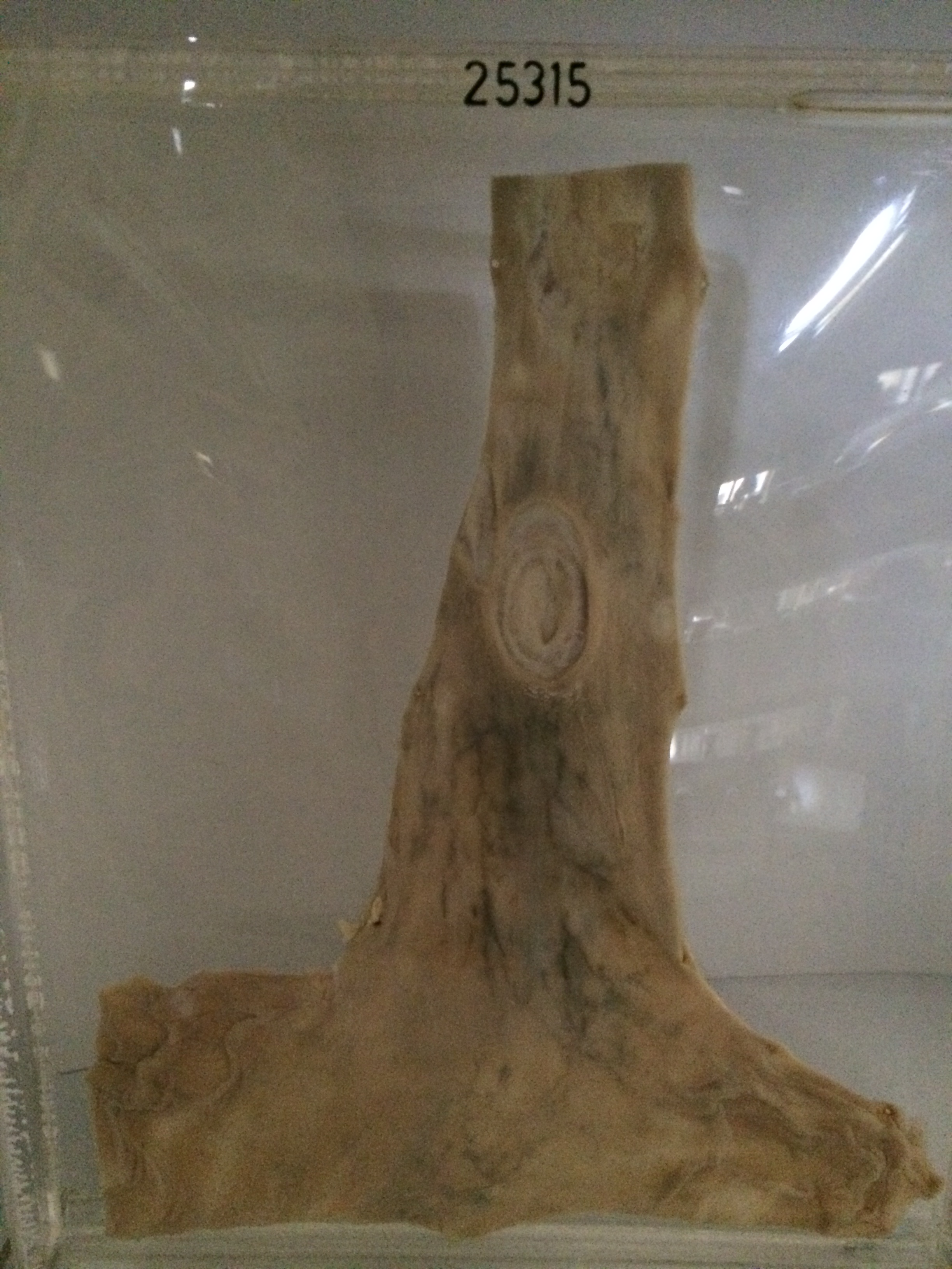
The specimen is the terminal 15 cms of the oesophagus and the cardiac end of the stomach. Eight cms above the cardio-oesophageal junction there is an indurated ovoid ulcer measuring 2.5 x 3 cms. The edge is regular and pale and the floor is indurated. The ulcer penetrates the muscle coat and a ring of sclerosed muscle can be seen in the base of the ulcer. There is a further scarred area 6 cms above the larger ulcer. This area shows two irregular shallow ulcers with serpiginous borders, and marked pale adjacent fibrosis.